Knee Muscle Anatomy Mri : Mri Knee Anatomy Knee Sagittal Anatomy Free Cross Sectional Anatomy Mri Knee Mri Anatomy. Mri for evaluating knee pain in older patients: Learn about knee anatomy muscle with free interactive flashcards. Click on the links to show each structure. 1 november 2002 mri anatomy of the knee and shoulder james y. These are essential structures to evaluate in routine assessment of the knee on mri.
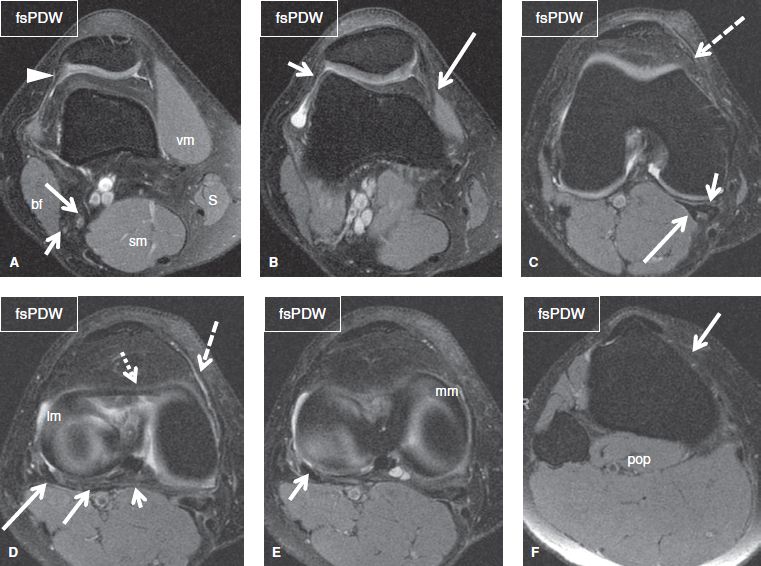
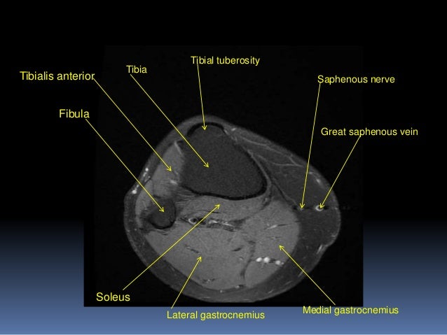
The journal of musculoskeletal medicine. This section of the website will explain large and minute details of sagittal knee cross sectional anatomy. Hurry, offer ends september 30. Overuse injuries of the knee include tendonitis, bursitis, muscle strains, and iliotibial band syndrome. Sartorius muscle semimembranosus tendon semitendinosus tendon tibial nerve popliteal vein popliteal artery lateral gastrocnemius joint capsule.
The Knee Musculoskeletal Key from musculoskeletalkey.com This section of the website will explain large and minute details of sagittal knee cross sectional anatomy. Want to learn more about it? Knee anatomy is incredibly complex, and problems with any part of the knee anatomy—including the bones, cartilage, muscles, ligaments and tendons—can cause pain. Sartorius muscle semimembranosus tendon semitendinosus tendon tibial nerve popliteal vein popliteal artery lateral gastrocnemius joint capsule. Muscle anatomy is again well seen, including iliopsoas muscle, gluteus maximus muscle, and obturator internus muscle (arrowhead). Learn anatomy using a full pacs! Our medical imaging anatomy course is now just $30! Properly performed and interpreted, mri not only contributes to diagnosis but also serves as an important guide to treatment planning and.
Seems like it should be pretty easy, right?
If the knee is flexed more than 5 degrees, it may appear lax. By now you probably know that the anatomy is deceptively complex, combinations of injuries can be challenging, and of course the referring clinician's expectations are as high as the range of meniscus injuries is wide. View of the anatomical labels. Each anatomical structure was labeled interactively. This mri knee sagittal cross sectional anatomy tool is. Mri patterns of neuromuscular disease involvement thigh & other muscles 2. Use the checklist to quiz yourself. Our medical imaging anatomy course is now just $30! A coronal scan goes through the knee, front. Click now to learn more about the bones, muscles, and soft tissues of these regions at leg and knee anatomy: To begin, we use a coronal scan of a left knee. 1 november 2002 mri anatomy of the knee and shoulder james y. Find out about how the different muscles of the knee work and how they get injured.
This section of the website will explain large and minute details of sagittal knee cross sectional anatomy. General anatomy and musculoskeletal system. Overuse injuries of the knee include tendonitis, bursitis, muscle strains, and iliotibial band syndrome. Mri for evaluating knee pain in older patients: View of the anatomical labels.
Kraeseriget from lh6.googleusercontent.com This is the only infrahyoid muscle not innervated by the ansa cervicalis, instead being supplied by fibres from the hypoglossal nerve. Knee pain that does not respond to treatment. The journal of musculoskeletal medicine. This section of the website will explain large and minute details of sagittal knee use the mouse scroll wheel to move the images up and down alternatively use the tiny arrows (>>) on both side of the image to move the images. Song, uc san francisco msiv gillian lieberman md. Hurry, offer ends september 30. Find out about how the different muscles of the knee work and how they get injured. 4, infrapatellar fat pad of hoffa.
Master leg and knee anatomy using our topic page.
Knee pain that does not respond to treatment. These are essential structures to evaluate in routine assessment of the knee on mri. Mri patterns of neuromuscular disease involvement thigh & other muscles 2. Mri for evaluating knee pain in older patients: If the knee is flexed more than 5 degrees, it may appear lax. Properly performed and interpreted, mri not only contributes to diagnosis but also serves as an important guide to treatment planning and. Magnetic resonance imaging (mri scan): Hurry, offer ends september 30. Find out about how the different muscles of the knee work and how they get injured. Learn anatomy using a full pacs! Musculoskeletal radiology south texas radiology group. Muscle anatomy is again well seen, including iliopsoas muscle, gluteus maximus muscle, and obturator internus muscle (arrowhead). Abnormal anatomy with normal signal.
To begin, we use a coronal scan of a left knee. A coronal scan goes through the knee, front. This mri knee cross sectional anatomy tool is absolutely free to use. Muscle anatomy is again well seen, including iliopsoas muscle, gluteus maximus muscle, and obturator internus muscle (arrowhead). Master leg and knee anatomy using our topic page.
Mri Knee Joint Anatomy from image.slidesharecdn.com Involved early gray = muscle: Magnetic resonance imaging (mri) interpretation of the knee is often a daunting challenge to the student or physician in training. 1 november 2002 mri anatomy of the knee and shoulder james y. Mr arthrogram knee loose osteochondral lesion. The main knee muscles are the quadriceps, hamstrings and calf muscles. Song, uc san francisco msiv gillian lieberman md. The muscles of the knee include the quadriceps, hamstrings, and the muscles of the calf. This mri knee sagittal cross sectional anatomy tool is.
General anatomy and musculoskeletal system.
General anatomy and musculoskeletal system. If the knee is flexed more than 5 degrees, it may appear lax. Related posts of knee muscle anatomy mri. Involved early gray = muscle: 4, infrapatellar fat pad of hoffa. The muscles of the knee include the quadriceps, hamstrings, and the muscles of the calf. Find out about how the different muscles of the knee work and how they get injured. Seems like it should be pretty easy, right? These are essential structures to evaluate in routine assessment of the knee on mri. Master leg and knee anatomy using our topic page. Knee muscles need to have both good strength and flexibility. Free cross sectional anatomy of the knee based on mri : Mr arthrogram knee loose osteochondral lesion.
Share :
Post a Comment
for "Knee Muscle Anatomy Mri : Mri Knee Anatomy Knee Sagittal Anatomy Free Cross Sectional Anatomy Mri Knee Mri Anatomy"


Post a Comment for "Knee Muscle Anatomy Mri : Mri Knee Anatomy Knee Sagittal Anatomy Free Cross Sectional Anatomy Mri Knee Mri Anatomy"